The Royal Netherlands Academy of Sciences, the Academical Medical Centre and the Technical University Delft cooperate in a joint effort to promote science by making image data, derived from previously made anatomical sections of human adult and embryonic orbits available to researchers.
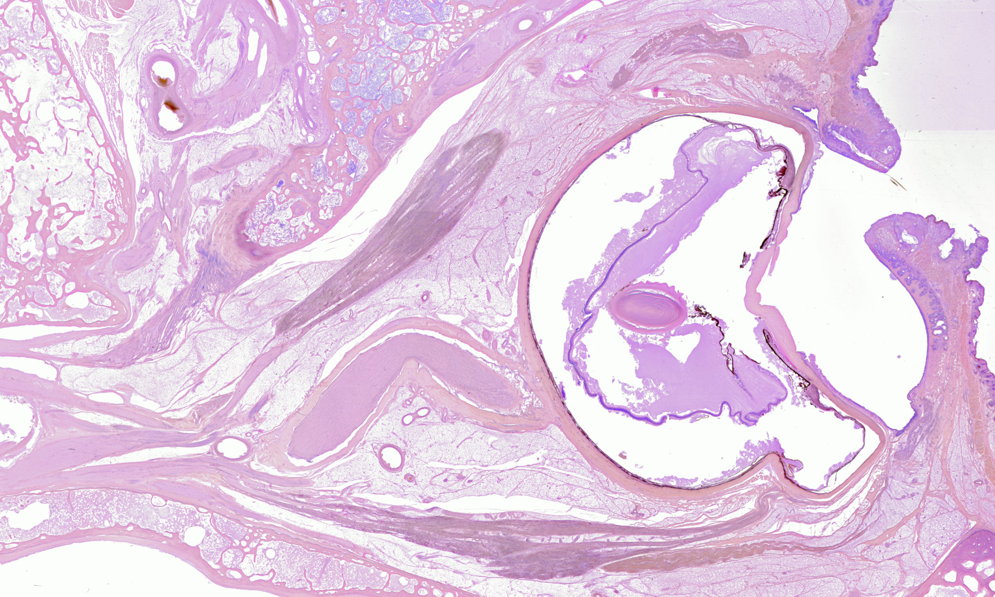
3000 Histological sections from 5 human adult orbits and 30 foetal heads and bodies form the heritage of the late Profs. A.A. Los & L. Koornneef, Dr. M.P. Bergen and Dr. A.B. de Haan at the Dept. of Anatomy of the Academical Medical Center and the Netherlands Ophthalmic Research Institute in Amsterdam. They comprise the largest and most detailed study of orbital anatomy ever. The hematoxylin-eosin and Giesson stains, however, have started to fade and nothing will remain in 10 or 20 years from now.
The celloidine sections, 60 100 m thick for adult and 25 50 m thick for embryonic sections have been scanned by Ben Willekens at high resolution (2500 dpi optically). These scans are now being converted by Joris van Swieten and Charl Botha into digital, high-resolution, 3-D reconstructions of the five orbits, accessible via the internet in the public domain. By making the 3-D reconstruction available in the public domain, many can join in the analysis of the anatomy of the orbit and of the way the eye is suspended in the orbit. The low resolution images can be downloaded from this site without obligation.
The final goal of the project is to offer a service whereby a researcher can choose any horizontal, sagittal, coronal or oblique plane in high-resolution 3-D bitmaps of the adult orbits and the embryonic heads and bodies, and order a recontructed, high-resolution section for download.
References
Bergen MP: Vasculatur architecture of the human orbit. PhD thesis, University of Amsterdam. Swets & Zeitlinger, 1982.
Koornneef L: Spatial aspects of orbital musculofribrous tissue in man. Ph.D. thesis. University of Amsterdam. Swets & Zeitlinger, 1976.