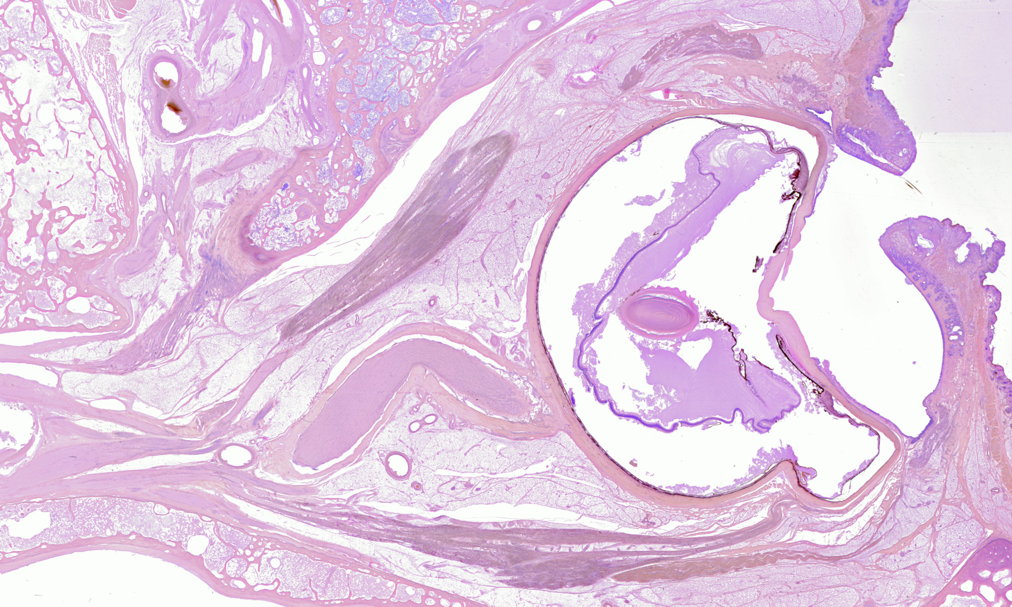
This movie shows a reconstruction of the head of a human fetus. The fetus is 79mm long, measured from crown to bottom (postovulatory age: between 13 and 15 weeks). It was cut along the frontal plane into 50 m thick sections, which were stained alternately with haematoxylin-eosin and haematoxylin-Van Gieson’s picro-fuchsin.
The reconstruction comprises sections #55 – #300. First, the original scans were downsampled to a resolution of 512×616 pixels. Secondly, the sections were segmented, i.e. the object of interest was separated from the background. Third, the RGB image data was converted to scalar image data using principal component analysis. Finally, reconstruction was performed by registration of pairs of adjacent images using normalised correlation as similarity measure. The centre of gravity of the segmentation masks was used to estimate an initial transform.
A number of interesting features can be recognised in the movie, for instance the cochlea, the vertebrae of the neck, and the teeth. These renderings should be interpreted with care, because they depend on the transfer function that was used, i.e. the mapping from data values to colour and transparency.
The reconstruction was done by Joris van Zwieten.
These sections were created, in the seventies, by Adriaan de Haan, anatomist and ophthalmologist working at The Netherlands Ophthalmic Research Institute.